CRISPR-Cas9
CRISPR gene editing technology that was the result of decades of research into the adaptive immune system of bacteria. In this exercise you will simulate the deletion of a gene using CRISPR-Cas9. You will first construct the pVG1 sgRNA expression vector from the pV1382 plasmid by cloning the appropriate sgRNA for deletion of the Saccharomyces cerevisiae ADE2 gene. The deletion will be carried out with a specific repair template DNA molcule. You will assemble each step using the ApE
You will retrieve the Saccharomyces cerevisiae ADE2 sequence, design the guide and repair template, and assemble a predicted plasmid in ApE. This exercise was adapted from the supplementary laboratory guide of [@vyas2019].
Background
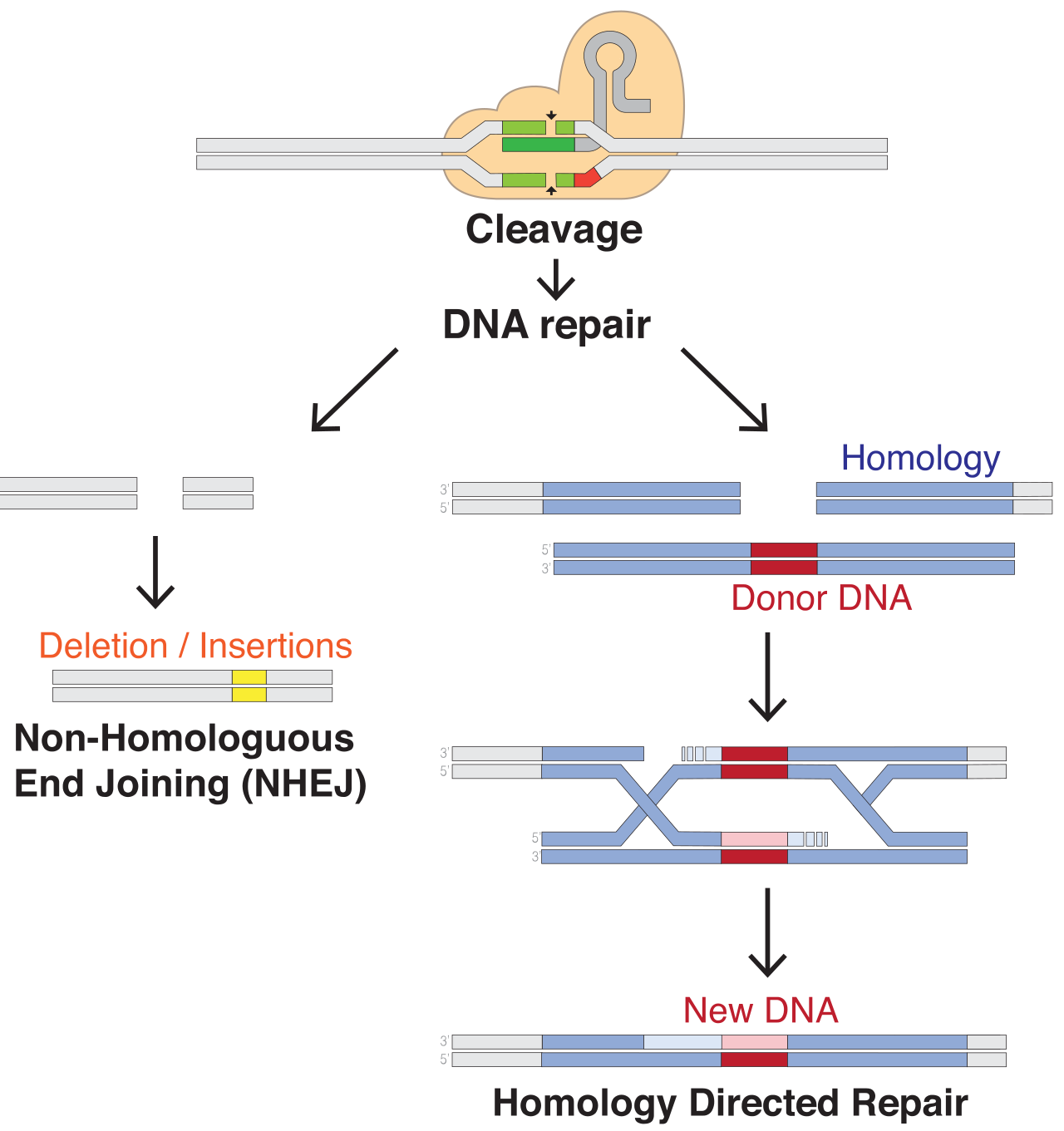
Mutagenesis can be random or targeted. CRISPR-Cas9 makes targeted mutagenesis possible by combining three components:
- Cas9, a nuclease that makes a double-strand break.
- A guide RNA (gRNA) containing a 20 nucleotide targeting sequence that directs Cas9 by base pairing.
- A repair template that can be copied by homologous recombination and thereby introduce a designed edit.
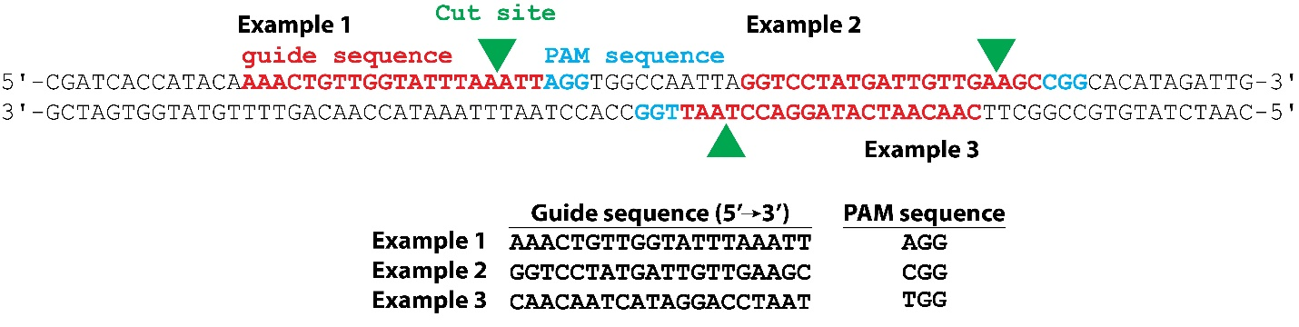
For the commonly used Streptococcus pyogenes Cas9 (SpCas9), a target must lie beside a 5’-NGG-3’ protospacer-adjacent motif (PAM). The PAM is part of the DNA target but is not part of the 20 nt guide sequence. Cas9 normally cleaves both strands three base pairs upstream of the PAM.
The repair template should alter the guide-binding sequence or PAM. Otherwise, Cas9 can recognize and cleave the repaired chromosome again.
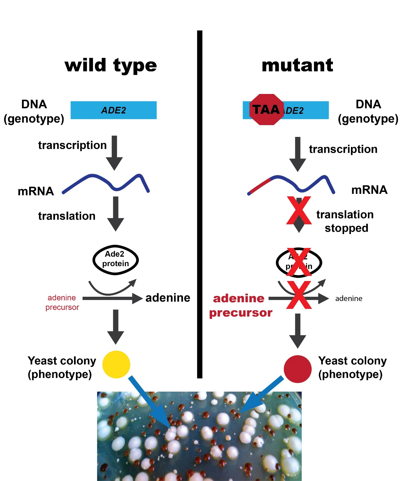
In S. cerevisiae, ADE2 encodes an enzyme in adenine biosynthesis. Loss of Ade2 activity causes a red pigment precursor to accumulate. This provides a convenient visible connection between a DNA edit and phenotype.
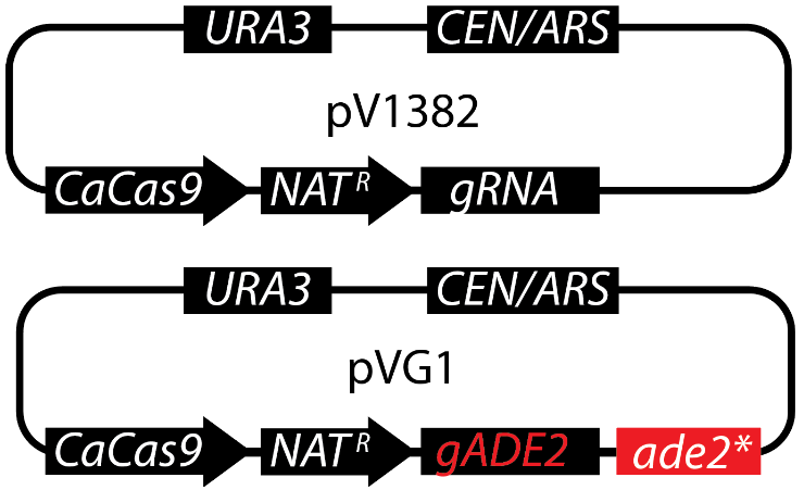
The parent plasmid pV1382 supplies Cas9, an sgRNA expression cassette, yeast replication and selection, and bacterial propagation functions. pVG1 adds an ADE2-specific guide (gADE2) and an ade2* repair template.
Software and sequence sources
Use ApE or another sequence editor that supports circular DNA, features, restriction analysis, reverse complementation, and pairwise alignment.
Retrieve the following records:
- SGD ADE2 / YOR128C: obtain the genomic DNA sequence, not only the translated protein.
- Plasmid sequence pV1382
cdseguid=EGjNBDMdbh_L18rl1eFSRc50Myc14965 bp, circular
IMPORTANT
The original sources document that pVG1 is pV1382 carrying
sgScADE2and an ADE2 stop-codon repair template, and Addgene records Gibson assembly as the cloning method. They do not provide a sufficiently detailed step-by-step historical construction in the teaching guide. The workflow below is therefore an in-silico reconstruction, checked against the deposited pVG1 sequence, rather than a claim about every experimental step used in the original laboratory.
Part 1 - Inspect the source records
- Download the ADE2 genomic sequence from SGD and open it in ApE.
- Confirm that the record is S. cerevisiae
YOR128Cand note the strand, chromosome coordinates, ORF length, and sequence version. - Mark the start and stop codons and translate the ORF. Confirm that the translation is uninterrupted in the wild-type sequence.
- Open pV1382 in a separate ApE window. Set the molecule to circular if the imported topology is not retained.
- Locate and annotate at least the following features:
CaCas9, the sgRNA cassette, the two BsmBI sites around its stuffer sequence,NATʳ,URA3,CEN/ARS, ampicillin resistance, and the bacterial origin.
Question 1: Why must the ADE2 DNA sequence, rather than only the Ade2 protein sequence, be used to design a guide?
Question 2: Which pV1382 features are needed in yeast, which are needed in E. coli, and which are directly responsible for genome editing?
Part 2 - Find the ADE2 target
A candidate target can occur on either DNA strand. On the displayed strand, search for NGG; the 20 bases immediately 5’ of each PAM form one class of candidates. To find candidates on the other strand, also search the reverse complement (equivalently, find CCN on the displayed strand).
- Make a table containing at least five ADE2 candidates.
- For each candidate record its 20 nt protospacer in the conventional 5’ to 3’ guide orientation, PAM, strand, position in the ORF, predicted cut position, and whether the cut is early enough to disrupt most of the protein.
- Locate the guide used in pVG1:
5'-ATTGGGACGTATGATTGTTG-3'- Find its PAM in the SGD sequence. Do not include the PAM in the guide insert.
- Mark the expected cleavage position three base pairs upstream of the PAM.
Question 3: On which strand is the published protospacer found, what is its PAM, and at which ADE2 coordinate is cleavage expected?
Question 4: What makes this target preferable or less preferable than the other candidates in your table? Consider position, specificity, and the ease of designing a repair template.
Part 3 - Simulate guide cloning into pV1382
pV1382 contains a removable stuffer flanked by BsmBI, a type IIS restriction enzyme. Because BsmBI cuts outside its recognition sequence, the recognition sites can disappear from the assembled product while defined overhangs direct insertion of the guide.
- Highlight the BsmBI sites in pV1382 and display the cut positions on both strands.
- Simulate BsmBI digestion and identify the backbone fragment containing
CaCas9,URA3,NATʳ, andCEN/ARS. - Design complementary oligonucleotides that place the 20 nt ADE2 protospacer between the BsmBI-generated ends. Derive the required overhangs from the actual pV1382 sequence rather than guessing them.
- Anneal the oligonucleotides in silico and insert the duplex into the cut backbone in the orientation that produces a functional sgRNA transcript.
- Name this intermediate
pV1382-gADE2and annotategADE2.
Question 5: Write both ordered oligonucleotides 5’ to 3’. Distinguish vector-compatible overhangs from the 20 nt sequence that becomes the guide.
Question 6: Which BsmBI sites are retained after assembly? Explain the result from the enzyme’s recognition and cleavage pattern.
Part 4 - Reconstruct the ade2* repair template
The pVG1 donor is homologous to the region surrounding the ADE2 cut but contains designed substitutions. The edit introduces premature stop codons and an EcoRI site. The relevant deposited donor sequence is:
ATGGATTCTAGAACAGTTGGTATATTAGGAGGGGGACAATTGGGACGTATGATTTTAATGAGAATTCGCAGCAAACAGGCTCAACATTAAGACGGTAATAC- Copy the corresponding wild-type interval from your SGD ADE2 record.
- Align the wild-type interval and donor sequence.
- Mark every mismatch and classify its effect as synonymous, missense, nonsense, PAM-disrupting, guide-disrupting, or restriction-site-forming. A change may have more than one role.
- Translate both aligned intervals in the ADE2 reading frame and mark the first premature stop.
- Confirm that
GAATTCcreates an EcoRI diagnostic site. - Explain which portions of the donor act as the left and right homology arms.
Question 7: Does the donor prevent renewed Cas9 cleavage by changing the PAM, the guide-binding region, or both?
Question 8: Why is an EcoRI site useful even though it is not required to abolish Ade2 function?
Question 9: Predict the phenotype of cells whose ADE2 locus copies this donor. Explain the complete chain from nucleotide change to colony colour.
Part 5 - Assemble a predicted pVG1
Addgene describes pVG1 as a Gibson-assembled derivative of pV1382. Gibson assembly requires overlapping ends and does not itself define the insertion position; that position must be inferred from the deposited sequences.
- Before opening pVG1, save a copy of
pV1382-gADE2. - Open the deposited pVG1 record only now.
- Align pV1382, your
pV1382-gADE2intermediate, and pVG1. Use the two pV1382/pVG1 junctions to locate the repair-template insertion site and determine its orientation. - In your saved intermediate, reproduce those junctions and insert the
ade2*donor in the inferred position and orientation. - Annotate the insert as
ade2* repair template, including its ADE2 homology and engineered EcoRI site. - Name the completed prediction
pVG1_reconstructedand save it as an annotated GenBank file.
Question 10: What sequence evidence identifies the two assembly junctions? Report enough bases on each side of each junction to make your answer unambiguous.
Question 11: Does sequence comparison support a single simultaneous assembly or a sequence of guide-cloning followed by donor insertion? State what can and cannot be concluded from the final plasmid sequence alone.
Part 6 - Validate the reconstruction
Do not validate only by looking at a circular map. Use independent checks.
- Compare the length and topology of
pVG1_reconstructedwith deposited pVG1. - Align the entire circular sequences. If their starting coordinates differ, rotate one sequence before interpreting the alignment.
- Verify both orientations because reverse-complemented circular records can describe the same physical molecule.
- Compare the positions and orientations of
gADE2,ade2*,CaCas9,URA3,NATʳ, andCEN/ARS. - Compare BsmBI and EcoRI restriction patterns.
- Calculate a circular DNA checksum if your available software supports one.
Complete the table:
| Check | Deposited pVG1 | pVG1_reconstructed | Match? |
|---|---|---|---|
| Length (bp) | |||
| Topology | circular | ||
| Number of BsmBI sites | |||
| Number of EcoRI sites | |||
gADE2 orientation | |||
ade2* orientation | |||
| Whole-sequence identity | reference | ||
| Circular checksum |
Question 12: If your sequence is not identical to the deposited pVG1, give the coordinates and sequence of every discrepancy. Classify each as a likely assembly error, record-orientation issue, uncertain historical junction, or source-version difference.
Submission
Submit:
pVG1_reconstructed.gb, with clear feature annotations;- your candidate-guide table;
- the wild-type/donor alignment with translated codons;
- the completed validation table;
- answers to Questions 1-12;
- a short cloning history describing each simulated operation and the evidence for it.
Learning objectives
After completing TP30, you should be able to:
- explain the roles of Cas9, a guide RNA, a PAM, and a repair template;
- find candidate SpCas9 targets on both strands of a gene;
- distinguish a protospacer from the adjacent PAM;
- predict the Cas9 cleavage position;
- design an edit that both changes a gene and prevents repeated cleavage;
- simulate type IIS cloning and sequence assembly in ApE;
- validate a reconstructed plasmid using size, restriction sites, annotations, and sequence comparison.
References and provenance
- Vyas VK, Bernstein DA. 2019. An introduction to CRISPR-mediated genome editing in fungi. Journal of Microbiology & Biology Education 20(3):20.3.62. https://doi.org/10.1128/jmbe.v20i3.1809
- Vyas VK, Bushkin GG, Bernstein DA, et al. 2018. New CRISPR mutagenesis strategies reveal variation in repair mechanisms among fungi. mSphere 3(2):e00154-18. https://doi.org/10.1128/mSphere.00154-18
- Addgene plasmid pV1382 #111436, RRID:Addgene_111436.
- Addgene plasmid pVG1 #111444, RRID:Addgene_111444.
- Saccharomyces Genome Database, ADE2/YOR128C.
Figures 1-4 were extracted without alteration from JMBE-20-62-s001.pdf, which is distributed with the article under the Creative Commons Attribution-NonCommercial-NoDerivatives 4.0 licence.
© Björn Johansson 2026