-
Pour 30–100 ml of dialysis buffer, usually ultra pure water or 1X TE (10 mM Tris-HCl, 1 mM EDTA, pH 8.0), into a petri plate or beaker.
-
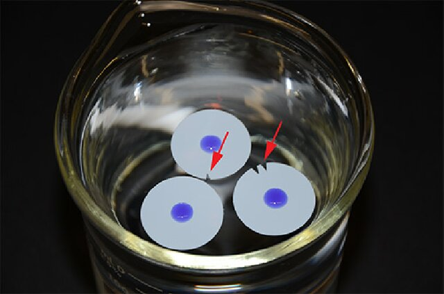
Float a 25 mm diameter, Type-VS Millipore membrane (MF type, VS filter, mean pore size = 0.025 μm, Millipore, Inc. vswp 02500) shiny side up on the dialysis buffer. Allow the floating filter to wet completely (~5 minutes) before proceeding. Make sure there are no air bubbles trapped under the filter. Mark the filters as indicated in the image below to identify different samples.
-
Pipette a few μl of the DNA droplet carefully onto the center of the filter. If the sample has too much phenol or chloroform, the drop will not remain in the center of the membrane and the dialysis should be discontinued until the organics are further removed. In most cases, this is performed by ethanol precipitation of the sample. If the test sample remains in the center of the membrane, pipette the remainder on to the membrane.
-
Cover the petri plate or beaker and dialyze for 1 to 4 hours. Do not allow the sample to flip or become covered with dialysis buffer.
-
Carefully retrieve the DNA droplet with a micro-pipette and place in a microcentrifuge tube. Rinse the spot on the membrane with 50 μl of 1X TE (10 mM Tris-HCl, 1 mM EDTA, pH 8.0) and add to the microcentrifuge tube.
-
Estimate the concentration of the DNA product using agarose gel electrophoresis or a spectrophotometer.
NOTE
Step 3 may be tricky for those with shaky hands or poor coordination. The filter has a tendency to move briskly around the surface as you touch it with the pipette tip. Practice with buffer droplets to master the technique before using a valuable sample.
Steps 2 to 4 can be repeated with fresh buffer or for longer times if additional dialysis is required.
Reference:
- Silhavy, T., Berman, M. and Enquist, L. Experiments with Gene Fusions, Cold Spring Harbor, N.Y. Press (1984).
- NEB